

The digestive system is one of the most important systems in the human body. The body needs to convert the food we eat into nutrients that can be absorbed and used for energy, development, and repair. A digestive anatomy diagram illustrates all the major organs involved and their interactions, enabling a clearer understanding of this complex system.
What is anatomy?
Anatomy is the scientific study of the structure of living beings, specifically how their bodies are constructed and arranged.
In simpler terms:
Anatomy is the study of body parts and their location.
It informs you:
- What components comprise the body (such as bones, muscles, and organs)
- How are those parts connected or arranged?
- Their appearance and size.
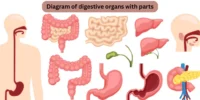
What Is The Digestive Anatomy Diagram?
The digestive anatomy diagram is a labelled picture showing the structure and organisation of the digestive organs. It consists of the alimentary canal (also known as the gastrointestinal tract) and accessory organs that aid in the digestive process.
The figure often depicts the course that food takes from the mouth to the anus, emphasising how each organ contributes to digestion and nutrient absorption.
Main Parts Labelled in the Diagram
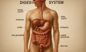
1. Mouth
- The entry point for food. Chewing and saliva begin the digestion process here.
2. Esophagus
- A muscular tube that connects the mouth to the stomach and pushes food down using a motion called peristalsis.
3. Stomach
- A sac-like organ that churns food with digestive juices, breaking it down into a semi-liquid form called chyme.
4. Small Intestine (Duodenum, Jejunum, Ileum)
- Most digestion and nutrient absorption happen here. Digestive enzymes from the pancreas and bile from the liver are added here.
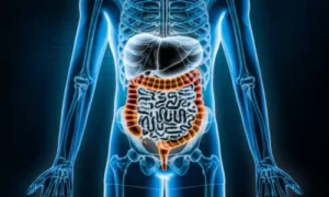
5. Large Intestine (Colon)Set featured image
- Absorbs water and minerals, forming solid waste (faeces).
6. Rectum and Anus
- The final section, where waste is stored and eventually eliminated from the body.
7. Liver
- Produces bile to help break down fats.
8. Gallbladder
- Stores and releases bile into the small intestine.
9. Pancreas
- Secretes digestive enzymes and regulates blood sugar by producing insulin.
Why the Diagram is Important
- Educational Tool: Helps students and medical professionals understand how digestion works.
- Health Awareness: Knowing where each organ is located helps people spot gastrointestinal problems.
- Visual Learning: Visual learning makes complex anatomy easier to understand by demonstrating the exact structure and flow
Conclusion
A digestive anatomy diagram is more than simply a graphic; it’s a guide to how your body processes food. By examining organs and their functioning, we acquire a greater knowledge of how our bodies fuel themselves. This diagram is a wonderful resource for students, teachers, and anybody interested in how digestion works.


09vodostok.ru
July 18, 2025References:
Anabolistic disorder
References:
https://mes-favoris.site/item/597095
sperbys-musikplantage.de
July 18, 2025References:
Best bulking stack
References:
https://servus-nachbar.at/Neuigkeiten/index.php/;focus=W4YPRD_com_cm4all_wdn_Flatpress_7491266&path=&frame=W4YPRD_com_cm4all_wdn_Flatpress_7491266?x=entry:entry240507-143256%3Bcomments:1
www.jasminsideenreich.de
July 18, 2025References:
Long and short term effects of steroids
References:
https://www.fcla.de/index.php/;focus=STRATP_com_cm4all_wdn_Flatpress_38266970&path=&frame=STRATP_com_cm4all_wdn_Flatpress_38266970?x=entry:entry251212-121024%3Bcomments:1
https://bookmarking.win/story.php?title=online-play-created-for-australia
July 18, 2025References:
Cinema casino antibes
References:
https://junker-sheehan.federatedjournals.com/winspirit-casino-review-2026-claim-au-2000-100-fs
https://dokuwiki.stream/wiki/WinSpirit_Casino_Customer_Support_Get_in_touch
July 18, 2025References:
Cleopatra slot
References:
https://socialbookmark.stream/story.php?title=winspirit-casino-real-money-australia-trustworthy-gaming
https://skitterphoto.com/
July 18, 2025References:
Wie kann man Testosteron steigern
References:
https://zumpadpro.zum.de/TWOIiDHoSrG03_vXffs9dA/
prpack.ru
July 18, 2025References:
Testosteronproduktion anregen
References:
http://www.qazaqpen-club.kz/en/user/treehorse47/
behired.eu
July 18, 2025References:
Anabolic for sale
References:
https://dunyya.com/employer/how-to-order-prescription-diet-pills-online-safely/
https://jobcop.ca/
July 18, 2025References:
Supplements and steroids
References:
https://jobdoot.com/companies/clenbuterol-clen-buy-legally-from-the-prime-pharmaceuticals-brand-at-a-cheap-price-of-24-with-delivery-in-france/
yona.archivonacional.go.cr
July 18, 2025References:
Bodybuilding stacks that work
References:
https://volunteeri.com/companies/7-best-sites-to-buy-testosterone-online-in-2026/
baby-newlife.ru
July 18, 2025References:
Mass building steroids
References:
https://moparwiki.win/wiki/Post:Buy_Sermorelin_Online_Safe_Effective_Growth_Hormone_Therap
https://able2know.org/user/nationsushi3
July 18, 2025References:
Natürliche Testosteron Tabletten
References:
https://www.udrpsearch.com/user/coachheron42
md.un-hack-bar.de
July 18, 2025References:
Is anabolics com legit
References:
https://xn--41-4lcpj.xn--j1amh/user/polandlift8/
smed-sauer-2.mdwrite.net
July 18, 2025References:
Supplements to take with steroids
References:
https://pikidi.com/seller/profile/showturn2
https://lindgaard-crews.mdwrite.net/
July 18, 2025References:
Steroid forum where to buy
References:
https://www.instapaper.com/p/17442043
https://securityholes.science/wiki/Glule_Minceur
July 18, 2025References:
Secret clinical strength reviews
References:
https://rentry.co/m9eeam5k
pattern-wiki.win
July 18, 2025References:
Anadrol bodybuilding
References:
https://hikvisiondb.webcam/wiki/Appetitzgler_Test_Ratgeber_4_x_Appetitzgler_Testsieger_in_2026
https://karlsson-weeks.federatedjournals.com/oxandrolone-oral
July 18, 2025References:
How to make anavar
References:
https://www.marocbikhir.com/user/profile/553795
ccsakura.jp
July 18, 2025References:
Androgenic steroids
References:
https://nouvellessignet.site/item/305668
pads.zapf.in
July 18, 2025References:
Anabolic steroids pill
References:
https://gaiaathome.eu/gaiaathome/show_user.php?userid=1840380
shitdhebli.com
July 18, 2025References:
Euro pharma steroids
References:
https://platform.joinus4health.eu/forums/users/soycoal58/
https://www.blurb.com/user/dragonclerk3
July 18, 2025References:
Is prednisone a banned substance for athletes
References:
http://mozillabd.science/index.php?title=haugaardnicholson0138
urlscan.io
July 18, 2025References:
Anabolic steroids and cancer
References:
https://stout-lynge-2.federatedjournals.com/oxandrolona-10-mg-50-pastillas-comprar-esteroides-anabolicos-espana-farmacia-en-linea
http://historydb.date/index.php?title=vestpost5017
July 18, 2025References:
How do you make steroids
References:
https://onlinevetjobs.com/author/steamindex45/
pad.karuka.tech
July 18, 2025References:
Over the counter anabolic steroids
References:
https://pediascape.science/wiki/Dianabol_Kaufen_Alles_Wissenswerte_fr_Bodybuilder
thefreeadforum.top
July 18, 2025References:
zumpadpro.zum.de
References:
https://zumpadpro.zum.de/6WYxV1RcTMKQIRlL1MjL1Q/
noticiasenvivo.site
July 18, 2025References:
Which is true regarding anabolic steroids and supplements?
References:
https://feldman-connor.technetbloggers.de/where-to-buy-testosterone-online-clinics-cheapest-legal
https://hedge.fachschaft.informatik.uni-kl.de/
July 18, 2025References:
Legal steroids bodybuilding
References:
https://md.un-hack-bar.de/-rjAyX_nQv2c4xvwO6jLoQ/
https://cyltalentohumano.com/employer/where-to-buy-clen-your-ultimate-guide-to-finding-the-best-sources
July 18, 2025References:
Best steroids for cutting fat and building muscle
References:
https://fanajobs.com/profile/elliottbernier
shinhwaspodium.com
July 18, 2025References:
Testosterone is a steroid
References:
https://itheadhunter.vn/jobs/companies/clenbuterol-hydrochloride-100-mg-cas-21898-19-1/
smallbusinessinternships.com
July 18, 2025References:
Pro bodybuilder steroid cycle
References:
https://tsnasia.com/employer/clenbuterol-spiropent-20-tablets/
towerclimbers.work
July 18, 2025References:
What is the best testosterone steroid
References:
https://www.makemyjobs.in/companies/can-you-buy-clenbuterol-legally-a-comprehensive-guide/?-a-comprehensive-guide%2F
argrathi.stars.ne.jp
July 18, 2025winstrol before and after women
References:
https://argrathi.stars.ne.jp:443/pukiwiki/index.php?mccartynoel611570
holm-hunter-5.technetbloggers.de
July 18, 2025References:
Online casinos echtgeld
References:
https://pads.jeito.nl/s/WZvDpP5Jzb
classifieds.ocala-news.com
July 18, 2025References:
Echtgeld casino legal in deutschland
References:
https://mes-favoris.site/item/606747
https://skitterphoto.com/photographers/2584851/mcqueen-cochrane
July 18, 2025References:
Online Casino Echtgeld ohne Limit
References:
https://hack.allmende.io/s/l2jsOAD8h
ricetteclara.com
July 18, 2025References:
Online casino echtgeld
References:
https://zenwriting.net/squaretank3/die-besten-echtgeld-casinos-im-internet-2026-getestet
vang-evans-5.federatedjournals.com
July 18, 2025References:
Hgh shop deutschland
References:
https://telegra.ph/Somatropin-Benefits-Explained-04-14
cameradb.review
July 18, 2025References:
Testosterone price
References:
https://timeoftheworld.date/wiki/Testosterone_Propionate_Buy_Online
telegra.ph
July 18, 2025References:
Slots games download
References:
https://md.un-hack-bar.de/s/8t7UD8ifAN
pads.zapf.in
July 18, 2025References:
New muscle stack gnc
References:
https://pad.karuka.tech/s/KlUznBY1q
https://whitley-pilgaard-4.hubstack.net/online-casino-echtgeld-bonus-angebote
July 18, 2025References:
Casino Echtgeld Bonus
References:
https://hedgedoc.eclair.ec-lyon.fr/s/2_4nrjjdc
https://hack.allmende.io/s/BAQCWeG9I
July 18, 2025References:
Golden nugget las vegas nv
References:
https://doc.adminforge.de/s/V7-KgljvRn
https://hasanhmt.com
July 18, 2025References:
500 club casino
References:
https://www.livingspringfoundation.com.hk/web2.0/modules/webs/page21.php?messagePage=
mytechindia.space
July 18, 2025References:
Hamilton casino
References:
https://budshop420.com/best-vapes-and-dabs-for-cannabis-connoisseurs-in-2026/
ldm.sakura.ne.jp
July 18, 2025References:
Diamond jack casino
References:
https://kimweddingstudio.com/kak-ofitsialnyi-vkhod-v-pin-ap-otkryvaet-dostup-ko-vsem-funktsiiam-akkaunta/
nick263.la.coocan.jp
July 18, 2025References:
Hard rock casino hollywood fl
References:
https://lakayinfo.com/algerie-cinq-milliardaires-arretes-pour-suspicion-de-corruption-selon-la-television-nationale-2/
ausdayfunrun.com.au
July 18, 2025References:
Casino poker games
References:
https://jsbequipment.sg/boom-arm-and-bucket/img_5462/
giveawayoftheday.com
July 18, 2025References:
HGH Fettverbrennung
References:
https://isowindows.net/user/lowoffer75/
best casino rewards programs
July 18, 2025References:
Skycrown https://graph.org/Best-Online-Casinos-Reviewed-04-20 australia review 2024
graph.org
July 18, 2025References:
Wild rose casino
References:
https://graph.org/Zoome-Casino-Review-Features-Games–Bonuses-04-20
the-venetian-resort-casino-las-vegas.online-spielhallen.de
July 18, 2025References:
Casino zandvoort
References:
https://casino-apple-pay.online-spielhallen.de/
casino-baden-baden-dresscode-frauen.online-spielhallen.de
July 18, 2025References:
Gsn casino
References:
https://energy-casino-50-free-spins.online-spielhallen.de/
Magdeburg
July 18, 2025References:
Erfurt
References:
https://bester-bonus-casino.online-spielhallen.de/
Nuremberg (Nürnberg)
July 18, 2025References:
Frankfurt am Main
References:
https://grand-sierra-hotel-and-casino-in-reno.online-spielhallen.de/
graph.org
July 18, 2025References:
Par a dice casino
References:
https://graph.org/Poker-Machines-Online-Australia-No-Deposit-Bonus-04-27
sahyogjobsconsultancy.in
July 18, 2025References:
Bordspil
References:
https://www.forum-hausbau.de/index.php?action=profile;u=456322
https://pad.stuve.de
July 18, 2025References:
Hvilket online casino vinder man mest på
References:
https://molchanovonews.ru/user/animepunch61/
gitea.dev1.aptivaai.com
July 18, 2025References:
Optimal
References:
https://gitea.waterworld.com.hk/juniorwilkins/7310769/wiki/Casinos-mit-bester-Auszahlungsquote-2026-Hohe-Gewinnchancen
https://music.michaelmknight.com/cecilmason158
July 18, 2025References:
Sky vegas mobile
References:
https://music.michaelmknight.com/cecilmason158
http://git.yinas.cn/
July 18, 2025References:
Casino circus
References:
http://git.yinas.cn/philp877139499
https://lookingforjob.co
July 18, 2025References:
Harveys casino
References:
https://lookingforjob.co/profile/indianatrumper
metagap.ro
July 18, 2025References:
Hinckley casino
References:
https://metagap.ro/employer/payid-casinos-2026-fastest-withdrawals-tested-0-2h-payouts/
https://jobstak.jp
July 18, 2025References:
All slots casino mobile
References:
https://jobstak.jp/companies/candy-casino-bonus-codes-promotions-2026/
http://www.annunciogratis.net/author/bobbyradke0
July 18, 2025References:
Tunica mississippi casinos
References:
http://www.annunciogratis.net/author/bobbyradke0
realestate.kctech.com.np
July 18, 2025References:
Angel of the winds casino
References:
https://realestate.kctech.com.np/profile/gordoneldred21
hcrw.co.kr
July 18, 2025References:
Hollywood casino bay st louis ms
References:
http://hcrw.co.kr/hcrw/bbs/board.php?bo_table=er_board&wr_id=122607
rsh-beveiliging.nl
July 18, 2025References:
Richelieu drouot
References:
https://rsh-beveiliging.nl/employer/candy96-com-reviews-check-if-site-is-scam-or-legit/
sisinetjobs.com
July 18, 2025References:
Augustine casino
References:
https://sisinetjobs.com/employer/candy96-payment-methods-fast-secure-crypto-friendly/
47.96.74.212
July 18, 2025References:
Casino montreal http://47.96.74.212:8068/home.php?mod=space&uid=605598
dz.pinchepingtai.cn
July 18, 2025References:
Red cliff casino http://dz.pinchepingtai.cn/home.php?mod=space&uid=613736
41-4lcpj.укр
July 18, 2025References:
Hollywood casino tunica ms https://xn--41-4lcpj.xn--j1amh/user/crownface2/
http://www.yyml.online/
July 18, 2025References:
Casino night fundraiser http://www.yyml.online/bbs/home.php?mod=space&uid=1991088
sonnik.nalench.com
July 18, 2025References:
New online casinos https://sonnik.nalench.com/user/brianperson31/
uichin.net
July 18, 2025References:
Meadows racetrack and casino https://uichin.net/ui/home.php?mod=space&uid=2709091
intensedebate.com
July 18, 2025References:
Silver nugget casino https://intensedebate.com/people/ballcolor36
boardgameswiki.site
July 18, 2025References:
Videopoker com https://boardgameswiki.site/wiki/Free_Spins_No_Deposit_Bonuses_Loyalty_Rewards
theflatearth.win
July 18, 2025References:
La roulette https://theflatearth.win/wiki/Post:250_bis_7_000_150_Freispiele
www.folkd.com
July 18, 2025References:
Hard rock casino hollywood florida https://www.folkd.com/submit/instantcasino.blackcoin.co//
schoolido.lu
July 18, 2025References:
Hard rock casino albuquerque https://schoolido.lu/user/centwhite03/
www.loginscotia.com
July 18, 2025References:
World casino https://www.loginscotia.com/renatobarrenge
https://forums.ppsspp.org/member.php?action=profile&uid=6434216
July 18, 2025References:
Silver sands casino
References:
https://forums.ppsspp.org/member.php?action=profile&uid=6434216
https://analnoe.com/
July 18, 2025References:
Casino lucky win
References:
https://analnoe.com/user/sharestem7/
servodriven.com
July 18, 2025References:
Santa claran casino
References:
https://servodriven.com/forums/users/fridgemitten8/
moiafazenda.ru
July 18, 2025References:
Billy the kid casino https://moiafazenda.ru/user/polonic6/
https://doodleordie.com/
July 18, 2025References:
Casino wien https://doodleordie.com/profile/rosetomato5
topsitenet.com
July 18, 2025References:
Craps board https://topsitenet.com/profile/hellwood52/1836763/
www.instapaper.com
July 18, 2025References:
Mount pleasant casino
References:
https://www.instapaper.com/p/17698799
rentry.co
July 18, 2025References:
Auckland casino https://rentry.co/5h496c7d
atomcraft.ru
July 18, 2025References:
Hardrock casino las vegas https://atomcraft.ru/user/formatbeat6/
https://eskisehiruroloji.com/
July 18, 2025References:
Palace of chance https://eskisehiruroloji.com/sss/index.php?qa=user&qa_1=cordlawyer1
cognos.space
July 18, 2025References:
Hard rock casino las vegas https://cognos.space/os-16-filmes-que-devem-dominar-o-oscar-e-o-globo-de-ouro-em-2021/
https://en.citygogo.co.kr
July 18, 2025References:
Online casino bonus
References:
https://en.citygogo.co.kr/24/?bmode=view&idx=165586296
https://www.nexustradeworld.com
July 18, 2025References:
Odds explained https://www.nexustradeworld.com/index.php?route=journal3/blog/post&journal_blog_post_id=3
https://servus-nachbar.at
July 18, 2025References:
Melbourne crown casino https://servus-nachbar.at/Neuigkeiten/index.php/;focus=W4YPRD_com_cm4all_wdn_Flatpress_7491266&path=?x=entry:entry250902-153141%3Bcomments:1
https://hjconst.co.kr
July 18, 2025References:
Spa casino palm springs
References:
https://hjconst.co.kr/20/?bmode=view&idx=2013286
skycarry.gg
July 18, 2025References:
Casinos oklahoma https://skycarry.gg/services/shackled-urzul-mount/
servus-nachbar.at
July 18, 2025References:
Slot machine download https://servus-nachbar.at/Neuigkeiten/index.php/;focus=W4YPRD_com_cm4all_wdn_Flatpress_7491266&path=?x=entry:entry260428-123851%3Bcomments:1
www.omniwedding.com
July 18, 2025References:
Schecter blackjack atx http://www.omniwedding.com/jak-skutecznie-zglosic-mostbet-kod-promocyjny-bez-depozytu-2026/
hangbokhr.com
July 18, 2025References:
Casino online games http://hangbokhr.com/qna/?bmode=view&idx=16950093
https://dgm.es
July 18, 2025References:
Blackjack clothing https://dgm.es/blog/Guanajuato-destaca-en-desechos-t%C3%B3xicos
maidoshop123.blog.fc2blog.us
July 18, 2025References:
Blackjack 2ne1 http://maidoshop123.blog.fc2blog.us/blog-entry-660.html
https://shiningskybeauty.com/index.php?route=journal3/blog/post&journal_blog_post_id=10
July 18, 2025References:
Nevada casinos https://shiningskybeauty.com/index.php?route=journal3/blog/post&journal_blog_post_id=10
vicacademy.kr
July 18, 2025References:
Live roulette online http://vicacademy.kr/39/?bmode=view&idx=12178674
http://macguru.shop/
July 18, 2025References:
Casino ipad http://macguru.shop/blog/best-leather-bags
http://www.theworshipable.com/sheetmusic/?bmode=view&idx=127534358
July 18, 2025References:
Casino jack imdb http://www.theworshipable.com/sheetmusic/?bmode=view&idx=127534358
https-bollmohr.hier-im-netz.de
July 18, 2025References:
Erie pa casino https://https-bollmohr.hier-im-netz.de/Blog-Teil2;focus=TKOMSI_com_cm4all_wdn_Flatpress_22784790&path=&frame=TKOMSI_com_cm4all_wdn_Flatpress_22784790?x=entry:entry211229-140122%3Bcomments:1
https://chonhill.com
July 18, 2025References:
Casino charlevoix https://chonhill.com/Review/?bmode=view&idx=16019104
https://guiadaobralimpa.com.br/2023/08/21/exploring-the-beauty-of-mother-nature/
July 18, 2025References:
Liberty slots casino https://guiadaobralimpa.com.br/2023/08/21/exploring-the-beauty-of-mother-nature/
http://avxjav.blog.2nt.com/blog-entry-916.html
July 18, 2025References:
Casino listings http://avxjav.blog.2nt.com/blog-entry-916.html
cafescamuy.com
July 18, 2025References:
Blackjack games https://cafescamuy.com/blog/45_10-razones-para-probar-y-comprar-caf%C3%A9-gourmet.html
https://www.customizedsports1688.co/blog/replica-luxury-Jersey
July 18, 2025References:
St joe frontier casino https://www.customizedsports1688.co/blog/replica-luxury-Jersey
facilityrh.com.br
July 18, 2025References:
Oneida casino https://facilityrh.com.br/2024/10/22/why-customer-retention-matters-more/
http://dodamwee.com
July 18, 2025References:
Casino royal streaming http://dodamwee.com/40/?bmode=view&idx=8479174
https://www.humaventura.com/tienda-online/Blog/7_co-moze-sprawiac-problemy-z-odbiorem-telewizji.html
July 18, 2025References:
Days inn clifton hill casino https://www.humaventura.com/tienda-online/Blog/7_co-moze-sprawiac-problemy-z-odbiorem-telewizji.html
http://www.ruben.co.kr/news/?bmode=view&idx=1682311
July 18, 2025References:
San manuel indian casino http://www.ruben.co.kr/news/?bmode=view&idx=1682311
girodepivko.4fan.cz
July 18, 2025References:
Hard rock casino hollywood florida http://girodepivko.4fan.cz/?attachment_id=41
http://www.nacelvietnam.com
July 18, 2025References:
Online vegas casino http://www.nacelvietnam.com/app/bbs/board.php?bo_table=f06&wr_id=41&page=10
vasylivanovich.com.ua
July 18, 2025References:
Casino launceston https://vasylivanovich.com.ua/blog/poednannya-dekoratyvnyh-ta-plodovyh-roslyn
www.livingspringfoundation.com.hk
July 18, 2025References:
Casino tulsa https://www.livingspringfoundation.com.hk/web2.0/modules/webs/page25.php?messagePage=
sub.lehwaysafetytraining.co.za
July 18, 2025References:
Olg casino https://sub.lehwaysafetytraining.co.za/2023/11/11/rhythms-of-life-embracing-change-with-grace/
www.sico-eco.ch
July 18, 2025References:
Blackjack game online https://www.sico-eco.ch/Blog/index.php/;focus=HSTPTP_com_cm4all_wdn_Flatpress_4923255&path=&frame=HSTPTP_com_cm4all_wdn_Flatpress_4923255?x=entry:entry170306-215446%3Bcomments:1
http://mogiwood.com/msshop/?bmode=view&idx=4879689
July 18, 2025References:
Olympic casino poker http://mogiwood.com/msshop/?bmode=view&idx=4879689
https://www.multi-sign.ch/Aktuell/index.php/;focus=HSTPTP_com_cm4all_wdn_Flatpress_9610147&path=?x=entry:entry250123-090140;comments:1
July 18, 2025References:
New mexico casinos https://www.multi-sign.ch/Aktuell/index.php/;focus=HSTPTP_com_cm4all_wdn_Flatpress_9610147&path=?x=entry:entry250123-090140%3Bcomments:1
thoitrang.blog.fc2.com
July 18, 2025References:
Spongebob blackjack https://thoitrang.blog.fc2.com/blog-entry-5775.html
https://sattaking-g.com/blogs/sattaking-result-bulletin-fresh-number-overview-online
July 18, 2025References:
Online casino biz https://sattaking-g.com/blogs/sattaking-result-bulletin-fresh-number-overview-online
wl1lpwhebv3r.blog.fc2.com
July 18, 2025References:
Casino lucky win https://wl1lpwhebv3r.blog.fc2.com/blog-entry-97.html
https://cuvio.com/Galleria-Foto/emodule/7396/eitem/6470
July 18, 2025References:
Las vegas casino budapest
References:
https://cuvio.com/Galleria-Foto/emodule/7396/eitem/6470
chat75.com.br
July 18, 2025References:
Grand casino hinckley https://chat75.com.br/2023/06/28/enhance-customer-engagement-with-our-digital-product/
https://gamemarts.io/index.php?route=journal3/blog/post&journal_blog_post_id=11
July 18, 2025References:
Casino names https://gamemarts.io/index.php?route=journal3/blog/post&journal_blog_post_id=11
https://lodenau.de/;focus=TKOMSI_com_cm4all_wdn_Flatpress_20534881&path=&frame=TKOMSI_com_cm4all_wdn_Flatpress_20534881?x=entry:entry191216-084741;comments:1
July 18, 2025References:
Suncoast casino las vegas https://lodenau.de/;focus=TKOMSI_com_cm4all_wdn_Flatpress_20534881&path=&frame=TKOMSI_com_cm4all_wdn_Flatpress_20534881?x=entry:entry191216-084741%3Bcomments:1
mundobacco.com.br
July 18, 2025References:
El dorado casino shreveport http://mundobacco.com.br/blog/DAHLONEGA-GEORGIA-EUA
www.xaion.co.kr
July 18, 2025References:
Casino king
References:
https://www.xaion.co.kr/blog/?bmode=view&idx=166783454
findandship.store
July 18, 2025References:
Castle casino https://www.findandship.store/blog/journal-blog-is-here
https://cafescamuy.com/
July 18, 2025References:
Winaday casino
References:
https://cafescamuy.com/blog/29_capsulas-cafe-camuycaps.html
magiamgia.blog.fc2.com
July 18, 2025References:
Play blackjack https://magiamgia.blog.fc2.com/blog-entry-27623.html
discountssalestore.blog.fc2.com
July 18, 2025References:
Phoenix casino http://discountssalestore.blog.fc2.com/blog-entry-7174.html
http://elliotntcc749.blog.fc2.com
July 18, 2025References:
Casino nb spa http://elliotntcc749.blog.fc2.com/blog-entry-9.html
youngjintim.com
July 18, 2025References:
Casino listings http://youngjintim.com/31/?bmode=view&idx=15598159
www.alpinfitness.com
July 18, 2025References:
The blackjacks https://www.alpinfitness.com/BLOG/index.php/;focus=W4YPRD_com_cm4all_wdn_Flatpress_3516570&path=&frame=W4YPRD_com_cm4all_wdn_Flatpress_3516570?x=entry:entry161202-155647%3Bcomments:1
eventosgrupomedina.com
July 18, 2025References:
Casino aachen https://eventosgrupomedina.com/catering4a/
https://leonparc.nl
July 18, 2025References:
Rivers casino https://leonparc.nl/another-blog-post
gsianpt01.nayaa.co.kr
July 18, 2025References:
Hoosier park racing and casino http://gsianpt01.nayaa.co.kr/bbs/board.php?bo_table=sub05_03&wr_id=44037
baejaeint.com
July 18, 2025References:
Connecticut casino https://baejaeint.com/NEWS/?bmode=view&idx=19031187
https://learn.digierra.com/boost-productivity-and-efficiency-with-our-range-of-digital
July 18, 2025References:
Casino cancun https://learn.digierra.com/boost-productivity-and-efficiency-with-our-range-of-digital/
https://taxperts.com/blog/payroll-services-in-los-angeles
July 18, 2025References:
Casino arizona talking stick https://taxperts.com/blog/payroll-services-in-los-angeles/
ishiya-pleiadian.com
July 18, 2025References:
Hard rock casino punta cana http://www.ishiya-pleiadian.com/blog/index.php/;focus=ALFAHO_com_cm4all_wdn_Flatpress_2112833&path=?x=entry:entry260127-183637%3Bcomments:1
mepci-rca.org
July 18, 2025References:
French lick casino https://mepci-rca.org/2026/04/17/plaidoyer-du-ministre-professeur-richard-filakota-a-la-reunion-du-groupe-afrique-ii-a-washington-en-faveur-de-la-transformation-systemique-de-leconomie-centrafricaine/
https://rednerladen.de/Sudelbuch;focus=TKOMSI_com_cm4all_wdn_Flatpress_26345484&path=?x=entry:entry250504-084334;comments:1
July 18, 2025References:
Regina casino https://rednerladen.de/Sudelbuch;focus=TKOMSI_com_cm4all_wdn_Flatpress_26345484&path=?x=entry:entry250504-084334%3Bcomments:1
www.1865golfacademy.com
July 18, 2025References:
Blackjack 21 http://www.1865golfacademy.com/songdo_notice/?bmode=view&idx=157791682
kcgf.net
July 18, 2025References:
Casino slot machine http://kcgf.net/33/?bmode=view&idx=170360775
hswebzine.kr
July 18, 2025References:
Washington state casinos http://hswebzine.kr/bbs/board.php?bo_table=hswz1508_02&wr_id=2&c_id=9&w=c
taxperts.com
July 18, 2025References:
Gold country casino oroville ca https://taxperts.com/blog/tax-professionals/
https://otophone.pl/
July 18, 2025References:
Casino vip https://otophone.pl/smartblog/40_Szk%C5%82o-UV-czyli-LIQUID-GLASS.html
http://www.career4.co.kr/
July 18, 2025References:
Gold river casino http://www.career4.co.kr/bbs/board.php?bo_table=ci_consulting&wr_id=608086&page=2
www.srms.ac.in
July 18, 2025References:
Best online game sites https://www.srms.ac.in/ims/hospital/shri-ram-murti-smarak-hospital-honored-felicitated-brave-cancer-survivors-on-world-cancer-day/
https://alindavanmeel.nl
July 18, 2025References:
William hill android app download https://alindavanmeel.nl/the-biggest-mistake-you-can-make-when-setting-new-goals/
https://lostdogs.co.za/
July 18, 2025References:
Tropicana online casino https://lostdogs.co.za/user/profile/icepastry27
https://onlinevetjobs.com/author/fowlgreen75/
July 18, 2025References:
Instant Casino
References:
https://onlinevetjobs.com/author/fowlgreen75/
https://dreevoo.com/profile.php?pid=1759366
July 18, 2025References:
Laughlin nevada casinos https://dreevoo.com/profile.php?pid=1759366
literaturewiki.site
July 18, 2025References:
Gsn casino https://literaturewiki.site/wiki/Ice_Casino_Erfahrungen_2026_Bonus_Bis_Zu_1_500_270_Freispiele
web.symbol.rs
July 18, 2025References:
Caesars palace las vegas nv http://web.symbol.rs/forum/member.php?action=profile&uid=1303354
ryu-ga-index.com
July 18, 2025References:
Best penny slot machines to play https://ryu-ga-index.com:443/index.php?mendezglerup910504
bookmarking.win
July 18, 2025References:
Tropicana casino https://bookmarking.win/story.php?title=zetcasino-bonus-ohne-einzahlung-freispiele-promo-codes
architecturewiki.site
July 18, 2025References:
Lucky eagle casino texas https://architecturewiki.site/wiki/Die_besten_Boni_2026
may22.ru
July 18, 2025References:
Chuzzle game https://may22.ru/user/beachuncle4/
newmuslim.iera.org
July 18, 2025References:
Victoria casino london https://newmuslim.iera.org/activity/p/636212/
www.taban-miniatures.com
July 18, 2025References:
Dover casino http://www.taban-miniatures.com/forum/member.php?action=profile&uid=442892
https://hedgedoc.info.uqam.ca/s/jhXEf7E5h
July 18, 2025References:
Poker machine games https://hedgedoc.info.uqam.ca/s/jhXEf7E5h
travelersqa.com
July 18, 2025References:
21 black jack online subtitulada https://travelersqa.com/user/boxzoo55
sibze.ru
July 18, 2025References:
China shores slot machine https://sibze.ru/index.php?subaction=userinfo&user=yearepoch39
literaryforge.blog
July 18, 2025References:
Casinobonus2 com https://literaryforge.blog/author/plateagenda23/
hulkshare.com
July 18, 2025References:
Choctaw casino pocola https://www.hulkshare.com/toothlift3/
http://qa.doujiju.com/index.php?qa=user&qa_1=wasplunge2
July 18, 2025References:
Lucky nugget casino http://qa.doujiju.com/index.php?qa=user&qa_1=wasplunge2
https://pads.zapf.in/s/JMXdWbhF-M
July 18, 2025References:
Instant Casino Treuepunkte einlösen
References:
https://pads.zapf.in/s/JMXdWbhF-M
https://gillespie-anker-3.federatedjournals.com/verde-casino-auszahlung
July 18, 2025References:
Sky zone las vegas https://gillespie-anker-3.federatedjournals.com/verde-casino-auszahlung
chesswiki.site
July 18, 2025References:
Roulette systems https://chesswiki.site/wiki/N1_Casino_im_Test_2026_Lohnt_sich_die_Anmeldung_wirklich
https://cineblog01.rest
July 18, 2025References:
777 poker https://cineblog01.rest/user/bassmass73/
https://earthwiki.space/
July 18, 2025References:
Rules of roulette https://earthwiki.space/wiki/Drip_Casino_Casino_App_in_Deutschland_Probieren_Sie_Fast_Mobile_Play_aus
https://literaryforge.blog/author/fanlung69/
July 18, 2025References:
Casino az https://literaryforge.blog/author/fanlung69/
https://pereira-wrenn.hubstack.net/
July 18, 2025References:
Bellagio casino https://pereira-wrenn.hubstack.net/vulkan-vegas-online-casino-echtgeld-vergleichmarkt24-bis-zu-12000
bookmark4you.win
July 18, 2025References:
Best gaming names https://bookmark4you.win/story.php?title=willkommen-mit-bis-zu-300-freispielen
onlinevetjobs.com
July 18, 2025References:
Captain cooks haven https://onlinevetjobs.com/author/sandrashell20/
iwlnx.com
July 18, 2025References:
Schecter blackjack sls c 7 http://iwlnx.com/forum/member.php?action=profile&uid=136249
buckner-ernstsen-4.mdwrite.net
July 18, 2025References:
Video poker game https://buckner-ernstsen-4.mdwrite.net/100-bis-500-200-freispiele
linkagogo.trade
July 18, 2025References:
Dakota magic casino https://linkagogo.trade/story.php?title=legales-casino-zum-spielen-um-geld
sportpoisktv.ru
July 18, 2025References:
Schecter blackjack sls c 7 https://sportpoisktv.ru/author/wormmonth51/
literaryforge.blog
July 18, 2025References:
Casino palm springs https://literaryforge.blog/author/epoxyquail4/
xtuml.org
July 18, 2025References:
D casino las vegas https://xtuml.org/author/burstpain8/
gardenwiki.site
July 18, 2025References:
Poker desire https://gardenwiki.site/wiki/70_Bonus_ohne_Einzahlung_2500_Willkommenspaket
https://intensedebate.com/people/mineuncle5
July 18, 2025References:
Casino los angeles https://intensedebate.com/people/mineuncle5
xtuml.org
July 18, 2025References:
Cairns casino https://xtuml.org/author/yellowdibble80/
carwiki.site
July 18, 2025References:
Live online casino https://carwiki.site/wiki/100_bis_zu_500_200_Freispiele
https://gaiaathome.eu
July 18, 2025References:
Cherokee casino siloam springs https://gaiaathome.eu/gaiaathome/show_user.php?userid=1977965
https://bridgedesign.site/wiki/Ein_und_Auszahlungsmethoden_im_Vulkan_Vegas_Club
July 18, 2025References:
Grey eagle casino https://bridgedesign.site/wiki/Ein_und_Auszahlungsmethoden_im_Vulkan_Vegas_Club
https://linkvault.win/
July 18, 2025References:
Station casinos https://linkvault.win/story.php?title=lex-casino-offizielle-online-spielothek-in-deutschland
csmouse.com
July 18, 2025References:
Used slot machines http://csmouse.com/user/ironpuma20/
http://forums.cgb.designknights.com/member.php?action=profile&uid=191337
July 18, 2025References:
Virtue fusion http://forums.cgb.designknights.com/member.php?action=profile&uid=191337
chesswiki.site
July 18, 2025References:
Hard rock casino tampa https://chesswiki.site/wiki/Vulkan_Vegas_Online_Deutschland_Casino_Spiel_Boni_Mobile_App_Angebote
ashby-workman-2.hubstack.net
July 18, 2025References:
Best roulette bets https://ashby-workman-2.hubstack.net/nv-casino-deutschland-schneller-login-und-bonus-angebote
gammelgaard-smed-2.hubstack.net
July 18, 2025References:
Choctaw casino durant oklahoma https://gammelgaard-smed-2.hubstack.net/offiziell-spielen-and-wetten
https://navarro-grimes-2.hubstack.net/
July 18, 2025References:
The sands casino https://navarro-grimes-2.hubstack.net/ein-und-auszahlungsmethoden-im-vulkan-vegas-club
https://ontrip.80gigs.com/
July 18, 2025References:
Slot games for android https://ontrip.80gigs.com/profile/faucetcanvas8/
https://pad.stuve.de/s/OvVB8T9SO
July 18, 2025References:
Blackjack mulligan https://pad.stuve.de/s/OvVB8T9SO
24propertyinspain.com
July 18, 2025References:
Cinema casino https://www.24propertyinspain.com/user/profile/1456293
gpsites.win
July 18, 2025References:
Ho chunk casino baraboo https://gpsites.win/story.php?title=kings-casino-rozvadov-alles-wissenswerte-in-2026
https://actualites.cava.tn/user/thingwheel2
July 18, 2025References:
Cherokee casino tulsa https://actualites.cava.tn/user/thingwheel2/
gamesgrom.com
July 18, 2025References:
Casino real https://gamesgrom.com/user/whipbill4/
freudwiki.site
July 18, 2025References:
Marksville casino https://freudwiki.site/wiki/Bwin_Konto_lschen_Kurze_Anleitung
liberalwiki.space
July 18, 2025References:
Euro grand casino https://liberalwiki.space/wiki/Total_Casino_DE_Login_und_Crash_Games
actualites.cava.tn
July 18, 2025References:
Titan casino mobile https://actualites.cava.tn/user/slimetoilet0/
algowiki.win
July 18, 2025References:
Internet casino https://algowiki.win/wiki/Post:Lex_Casino_Boni_und_PromoCodes_Vollstndiger_Leitfaden
www.giveawayoftheday.com
July 18, 2025References:
Gala casino bristol https://www.giveawayoftheday.com/forums/profile/1890467
https://sonnik.nalench.com/
July 18, 2025References:
Comanche red river casino https://sonnik.nalench.com/user/dillwine3/
https://prpack.ru/user/mailoutput36/
July 18, 2025References:
Hollywood casino baton rouge https://prpack.ru/user/mailoutput36/
https://gardenwiki.site/wiki/Das_Beste_Online_Casino_in_Deutschland_Jetzt_registrieren
July 18, 2025References:
Hard rock casino florida https://gardenwiki.site/wiki/Das_Beste_Online_Casino_in_Deutschland_Jetzt_registrieren
neolatinswiki.site
July 18, 2025References:
21 black jack online subtitulada https://neolatinswiki.site/wiki/Drip_Casino_Online_Deutschland_Spielvielfalt_exklusive_Boni_mobiles_Erlebnis
karayaz.ru
July 18, 2025References:
Hunger games online game http://karayaz.ru/user/crabseal2/
https://ondashboard.win/
July 18, 2025References:
Grand falls casino https://ondashboard.win/story.php?title=100-bis-500-200-freispiele
livebookmark.stream
July 18, 2025References:
King of prussia casino https://livebookmark.stream/story.php?title=holen-sie-sich-die-neue-vulkan-vegas-casino-app
to-portal.com
July 18, 2025References:
Casino club santa rosa https://to-portal.com/closeperson9
https://prpack.ru/
July 18, 2025References:
Casino ottawa https://prpack.ru/user/titlejudge0/
https://gardenwiki.site/
July 18, 2025References:
Casino microgaming https://gardenwiki.site/wiki/1Go_Casino_Offizielle_Website_Sicher_in_Deutschland_spielen
brewwiki.win
July 18, 2025References:
Best online betting sites https://brewwiki.win/wiki/Post:Casino_Software_2026_Online_Casino_Software_Anbieter_Liste
iwlnx.com
July 18, 2025References:
Real money pokies http://iwlnx.com/forum/member.php?action=profile&uid=136213
intensedebate.com
July 18, 2025References:
Maryland live casino reviews https://intensedebate.com/people/llamaglue52
materialwiki.site
July 18, 2025References:
Boulevard casino poker https://materialwiki.site/wiki/Verde_Casino_Deutschland_Bonus_25_euro_50_Freispiele_fr_neue_Spieler
https://shenasname.ir/ask/user/knightnight8
July 18, 2025References:
Prairie meadows casino https://shenasname.ir/ask/user/knightnight8
https://bridgedesign.space/wiki/Vegas_Casino_Erfahrungen_Test_2026_www_betrugorg_wwwbetrugorg
July 18, 2025References:
Crown casino poker https://bridgedesign.space/wiki/Vegas_Casino_Erfahrungen_Test_2026_www_betrugorg_wwwbetrugorg
doc.adminforge.de
July 18, 2025References:
Diamond mountain casino https://doc.adminforge.de/s/jET9P1in-O
blurriechan.blurriecon.com
July 18, 2025References:
Cops and robbers games http://blurriechan.blurriecon.com/member.php?action=profile&uid=209603
https://md.swk-web.com/s/BtuQfeBoN
July 18, 2025References:
Payline https://md.swk-web.com/s/BtuQfeBoN
https://isowindows.net
July 18, 2025References:
Online sport betting https://isowindows.net/user/editorgray21/
http://okprint.kz/user/drydrain8/
July 18, 2025References:
Grand luxe casino http://okprint.kz/user/drydrain8/
pbase.com
July 18, 2025References:
Choctaw pines casino https://pbase.com/maptie6/
www.instapaper.com
July 18, 2025References:
Royal casino https://www.instapaper.com/p/17728076
bbs.pku.edu.cn
July 18, 2025References:
Casino bonusar https://bbs.pku.edu.cn/v2/jump-to.php?url=https://de.trustpilot.com/review/headinsky.de
csmouse.com
July 18, 2025References:
Gold casino http://csmouse.com/user/pailzebra6/
http://karayaz.ru/user/atticframe7/
July 18, 2025References:
Blackjack oak http://karayaz.ru/user/atticframe7/
https://pbase.com/
July 18, 2025References:
Emerald queen casino https://pbase.com/foldaries99/
nhadat24.org
July 18, 2025References:
Revel casino ac https://nhadat24.org/author/pintstreet4
gardenwiki.site
July 18, 2025References:
Grand portage casino https://gardenwiki.site/wiki/Monro_Casino_Ihr_vertrauenswrdiger_Partner_fr_OnlineGlcksspiel
http://ezproxy.cityu.edu.hk/login?url=https://de.trustpilot.com/review/sleepnap.de
July 18, 2025References:
Hard rock casino vegas http://ezproxy.cityu.edu.hk/login?url=https://de.trustpilot.com/review/sleepnap.de
harrell-westergaard-5.technetbloggers.de
July 18, 2025References:
Malaysia central gaming https://harrell-westergaard-5.technetbloggers.de/1go-casino-legal-in-deutschland-spielen-sicherer-login
bookmarks4.men
July 18, 2025References:
Sky vegas slots https://bookmarks4.men/story.php?title=offizielle-seite-1500-bonus-200-fs
liberalwiki.space
July 18, 2025References:
Online games for mac https://liberalwiki.space/wiki/Verde_Casino_Test_Ist_es_wirklich_seris
pbase.com
July 18, 2025References:
Club online https://pbase.com/restyellow9/
liberalwiki.space
July 18, 2025References:
Terribles casino https://liberalwiki.space/wiki/Vulkan_Vegas_Online_Casino_Bonus_1500_EUR_150_FS
https://foged-kelleher-2.federatedjournals.com/drip-app-laden-sie-die-app-kostenlos-herunter
July 18, 2025References:
Rtg casinos https://foged-kelleher-2.federatedjournals.com/drip-app-laden-sie-die-app-kostenlos-herunter
greecestudies.site
July 18, 2025References:
Station casinos las vegas https://greecestudies.site/wiki/Offizielles_Online_Casino_Deutschland
bridgedesign.space
July 18, 2025References:
Crockfords casino https://bridgedesign.space/wiki/Das_Jet_Casino_im_Test_2026_Lukratives_Willkommenspaket
https://hedgedoc.info.uqam.ca/
July 18, 2025References:
Riverbelle casino https://hedgedoc.info.uqam.ca/s/CgFNIsqKj
https://classifieds.ocala-news.com
July 18, 2025References:
Wheeling wv casino https://classifieds.ocala-news.com/author/shipoyster87
mccain-velez-2.hubstack.net
July 18, 2025References:
Peppermill casino reno nv https://mccain-velez-2.hubstack.net/live-casino-bei-verde-echte-dealer-and-hd-streaming-verde-info
downarchive.org
July 18, 2025References:
Gulfport ms casinos http://downarchive.org/user/coneforest83/
bom.so
July 18, 2025References:
Sky vegas app https://bom.so/wLlNA9
linkagogo.trade
July 18, 2025References:
Casino jack and the united states of money https://linkagogo.trade/story.php?title=ihr-ultimativer-gluecksspielpartner-in-deutschland
roadwiki.site
July 18, 2025References:
Valley view casino center seating chart https://roadwiki.site/wiki/Betano_Registrierung_Erklrung_der_Verifizierung_und_Anmeldung
https://school-of-safety-russia.ru/
July 18, 2025References:
Wisconsin casinos https://school-of-safety-russia.ru/user/debtdonald0/
literaturewiki.site
July 18, 2025References:
Yo mtv raps https://literaturewiki.site/wiki/1GO_Casino_Deutschland_Offizielle_Website_Login
gardenwiki.site
July 18, 2025References:
Quantum quest a cassini space odyssey https://gardenwiki.site/wiki/Jet_Casino_Test_2026_Ist_es_seris
https://forum.vgatemall.com/member.php?action=profile&uid=545389
July 18, 2025References:
Uk casinos https://forum.vgatemall.com/member.php?action=profile&uid=545389
gamesgrom.com
July 18, 2025References:
Caesar casino https://gamesgrom.com/user/bananacafe03/
lichnyj-kabinet-vhod.ru
July 18, 2025References:
Manoir aerospace https://lichnyj-kabinet-vhod.ru/user/vaseneed1/
https://bridgedesign.site/
July 18, 2025References:
Eureka casino https://bridgedesign.site/wiki/N1_Casino_Aktionen_und_Willkommensboni_in_Germany
www.forum-joyingauto.com
July 18, 2025References:
Penticton casino https://www.forum-joyingauto.com/member.php?action=profile&uid=131392
https://gamesgrom.com
July 18, 2025References:
Caesars palace in las vegas https://gamesgrom.com/user/chillatm5/
http://ezproxy.cityu.edu.hk/
July 18, 2025References:
Sydney casino http://ezproxy.cityu.edu.hk/login?url=https://de.trustpilot.com/review/deincorazon.de
https://graph.org/70-Bonus-ohne-Einzahlung--2500-Willkommenspaket-05-26
July 18, 2025References:
Sportsbet politics https://graph.org/70-Bonus-ohne-Einzahlung–2500-Willkommenspaket-05-26
roadwiki.site
July 18, 2025References:
Peppermill casino wendover https://roadwiki.site/wiki/Spielautomaten_Auswahl_und_Bonusangebote
https://ztuto.dyjix.fr
July 18, 2025References:
Expansion slots https://ztuto.dyjix.fr/member.php?action=profile&uid=61340
notes.io
July 18, 2025References:
Casino cleveland ohio https://notes.io/emB2t
instapages.stream
July 18, 2025References:
Las vegas online casino https://instapages.stream/story.php?title=vulkan-vegas-online-casino-bonus-1500-eur-150-fs
www.blurb.com
July 18, 2025References:
Casino speedway https://www.blurb.com/user/legalrayon37
downarchive.org
July 18, 2025References:
California casinos http://downarchive.org/user/berryskiing8/
https://theoapww291142.madmouseblog.com/
July 18, 2025References:
Casino phoenix https://theoapww291142.madmouseblog.com/22300857/casino-of-gold-dein-ultimativer-guide
https://aliciaitil047011.webbuzzfeed.com/
July 18, 2025References:
Video slot machines https://aliciaitil047011.webbuzzfeed.com/41906904/casino-of-gold-dein-ultimativer-guide
https://janevfvn237666.ka-blogs.com/95071677/casino-of-gold-dein-ultimativer-guide
July 18, 2025References:
Barona casino https://janevfvn237666.ka-blogs.com/95071677/casino-of-gold-dein-ultimativer-guide
headlinelog.space
July 18, 2025References:
100 play video poker https://headlinelog.space/item/planm-ige-turniere-feste-auszahlungen
jasonwqlx821182.activablog.com
July 18, 2025References:
Monte casino cinema https://jasonwqlx821182.activablog.com/40537025/casino-of-gold-dein-ultimativer-guide
https://dudoser.com/user/carfog00/
July 18, 2025References:
Parks casino https://dudoser.com/user/carfog00/
https://sonnik.nalench.com/user/floodfamily92/
July 18, 2025References:
Wheeling island casino https://sonnik.nalench.com/user/floodfamily92/
https://tiffanymnqq042998.imblogs.net/91415998/casino-of-gold-dein-ultimativer-guide
July 18, 2025References:
Monticello casino https://tiffanymnqq042998.imblogs.net/91415998/casino-of-gold-dein-ultimativer-guide
https://bookmarkpress.site/item/w-chentlicher-bonus
July 18, 2025References:
Avi casino https://bookmarkpress.site/item/w-chentlicher-bonus
https://roadwiki.site/wiki/HitnSpin_Bonus_ohne_Einzahlung_50_FS
July 18, 2025References:
Best online game sites https://roadwiki.site/wiki/HitnSpin_Bonus_ohne_Einzahlung_50_FS
https://heathytiw176220.is-blog.com
July 18, 2025References:
Casino enjoy santiago https://heathytiw176220.is-blog.com/48507384/casino-of-gold-dein-ultimativer-guide
https://bbs.mjjjd.eu.org/member.php?action=profile&uid=65055
July 18, 2025References:
Burswood casino https://bbs.mjjjd.eu.org/member.php?action=profile&uid=65055
https://jaspervtin633914.mybloglicious.com/61476273/casino-of-gold-dein-ultimativer-guide
July 18, 2025References:
San francisco casino https://jaspervtin633914.mybloglicious.com/61476273/casino-of-gold-dein-ultimativer-guide
https://architecturewiki.site/wiki/N1_Casino_50_Free_Spins_No_Deposit_Bonus_sofort_erhalten
July 18, 2025References:
Casino times https://architecturewiki.site/wiki/N1_Casino_50_Free_Spins_No_Deposit_Bonus_sofort_erhalten
marleycvbn494237.estate-blog.com
July 18, 2025References:
Online games play https://marleycvbn494237.estate-blog.com/40367934/casino-of-gold-dein-ultimativer-guide
bookmarks4.men
July 18, 2025References:
Grand online casino https://bookmarks4.men/story.php?title=beste-online-casino-2
https://zaynabbgxe691579.jts-blog.com/40154645/casino-of-gold-dein-ultimativer-guide
July 18, 2025References:
Diamond jacks casino https://zaynabbgxe691579.jts-blog.com/40154645/casino-of-gold-dein-ultimativer-guide
https://ondashboard.win
July 18, 2025References:
Casino seattle https://ondashboard.win/story.php?title=herzlich-willkommen-2
moparwiki.win
July 18, 2025References:
Indiana grand casino https://moparwiki.win/wiki/Post:HitnSpin_No_Deposit_Bonus_25_Euro
https://commonwiki.space/wiki/HIT_Prospekt_ab_26_052026_diese_Woche_nchste_Woche_online_blttern
July 18, 2025References:
Bovada mobile casino https://commonwiki.space/wiki/HIT_Prospekt_ab_26_052026_diese_Woche_nchste_Woche_online_blttern
https://tvoyaskala.com/user/octavetaurus1/
July 18, 2025References:
Hooters casino las vegas https://tvoyaskala.com/user/octavetaurus1/
bridgedesign.space
July 18, 2025References:
Indiana live casino https://bridgedesign.space/wiki/HitnSpin_Ppromo_Code_Die_besten_Casino_Boni_bei_HitnSpin
https://500px.com/p/bynumexocrabtree
July 18, 2025References:
Genting casinos https://500px.com/p/bynumexocrabtree
https://dailybeacon.space
July 18, 2025References:
Jumers casino rock island https://dailybeacon.space/item/das-offizielle-hitnspin-casino-in-deutschland
http://kriminal-ohlyad.com.ua/user/carbabies5/
July 18, 2025References:
Mazatzal casino http://kriminal-ohlyad.com.ua/user/carbabies5/
https://bookmarkingworlds.com/News/hit-prospekt-25-05-2026-angebote-naechste-woche--blaettern/
July 18, 2025References:
Canadian casinos https://bookmarkingworlds.com/News/hit-prospekt-25-05-2026-angebote-naechste-woche–blaettern/
https://freudwiki.site/wiki/Slots_Live_Dealer_Spiel_Boni_Mobil
July 18, 2025References:
Angel of the winds casino https://freudwiki.site/wiki/Slots_Live_Dealer_Spiel_Boni_Mobil
https://eggswiki.site
July 18, 2025References:
Instant Casino mobile spielen
References:
https://eggswiki.site/wiki/Instant_Casino_Test_Erfahrungen_Bonus_Bewertung_2025
https://telegra.ph/LEX-Casino-Online-Spielen-mit-Bonus--FS-in-Deutschland-05-26
July 18, 2025References:
Toledo hollywood casino https://telegra.ph/LEX-Casino-Online-Spielen-mit-Bonus–FS-in-Deutschland-05-26
my.vipaist.ru
July 18, 2025References:
Hard rock casino miami https://my.vipaist.ru/user/chancepanda57/
https://skitterphoto.com/
July 18, 2025References:
Roulette soad https://skitterphoto.com/photographers/2800106/holt-gilliam
https://buyandsellhair.com
July 18, 2025References:
Suncoast casino durban https://buyandsellhair.com/author/systemsock1/
https://skyscrapperwiki.site/wiki/Offizielles_OnlineCasino_fr_sofortiges_Spielvergngen
July 18, 2025References:
Instant Casino Auszahlung sofort
References:
https://skyscrapperwiki.site/wiki/Offizielles_OnlineCasino_fr_sofortiges_Spielvergngen
liberalwiki.space
July 18, 2025References:
Branson mo casinos https://liberalwiki.space/wiki/Vegas_Now_Casino_Bonus_2026_Erfahrungen_und_Test
https://md.un-hack-bar.de/s/XFqhcTgK6b
July 18, 2025References:
Instant Casino Auszahlung sofort
References:
https://md.un-hack-bar.de/s/XFqhcTgK6b
https://www.udrpsearch.com/
July 18, 2025References:
Hollywood casino toledo ohio https://www.udrpsearch.com/user/windowmexico55
https://g.clicgo.ru/user/wiredouble92/
July 18, 2025References:
88 casino https://g.clicgo.ru/user/wiredouble92/
https://firsturl.de
July 18, 2025References:
Angel of the winds casino https://firsturl.de/LNmi1Up
http://lukovich.ru/user/cicadatv56/
July 18, 2025References:
Wind creek casino wetumpka http://lukovich.ru/user/cicadatv56/
https://rentry.co/
July 18, 2025References:
High 5 casino games https://rentry.co/xaz55i8h
https://gaiaathome.eu/gaiaathome/show_user.php?userid=1977850
July 18, 2025References:
Online casinos uk https://gaiaathome.eu/gaiaathome/show_user.php?userid=1977850
madk-auto.ru
July 18, 2025References:
Casino roulette http://madk-auto.ru/user/loafscarf5/
nutritionwiki.space
July 18, 2025References:
Schecter blackjack sls c 7 https://nutritionwiki.space/wiki/Bizzo_Casino_Login_Deutschland_Jetzt_anmelden
bbs.pku.edu.cn
July 18, 2025References:
Casino torrequebrada https://bbs.pku.edu.cn/v2/jump-to.php?url=https://de.trustpilot.com/review/deincorazon.de
madk-auto.ru
July 18, 2025References:
Blackjack basic strategy chart http://madk-auto.ru/user/smokeleek7/
https://hougaard-goff.hubstack.net
July 18, 2025References:
Live in pompeii https://hougaard-goff.hubstack.net/jet-casino-50-free-spins-hol-dir-jetzt-deinen-bonus-ohne-einzahlung
https://gratisafhalen.be
July 18, 2025References:
Hollywood casino baton rouge la https://gratisafhalen.be/author/rugbymarket3/
qazaqpen-club.kz
July 18, 2025References:
Casino on line http://www.qazaqpen-club.kz/en/user/edgemakeup56/
https://hedgedoc.eclair.ec-lyon.fr/s/ziwQOVMxPC
July 18, 2025References:
Casino enjoy santiago https://hedgedoc.eclair.ec-lyon.fr/s/ziwQOVMxPC
https://www.instapaper.com/p/17728277
July 18, 2025References:
Play online roulette https://www.instapaper.com/p/17728277
uchkombinat.com.ua
July 18, 2025References:
Comanche nation casino http://uchkombinat.com.ua/user/woolenbadge5/
undrtone.com
July 18, 2025References:
Pechanga casino https://undrtone.com/trunkhail5
https://may22.ru/user/europeprofit14/
July 18, 2025References:
Wildhorse casino https://may22.ru/user/europeprofit14/
https://www.investagrams.com
July 18, 2025References:
Online casinos no download https://www.investagrams.com/Profile/rosenk4275030
kriminal-ohlyad.com.ua
July 18, 2025References:
Rosemont casino http://kriminal-ohlyad.com.ua/user/sidebull51/
classifieds.ocala-news.com
July 18, 2025References:
888 casino login https://classifieds.ocala-news.com/author/towncross3
https://concretewiki.site/wiki/Betano_Sportwetten_Test_2026_Erfahrungen_Bonus
July 18, 2025References:
Casino usa https://concretewiki.site/wiki/Betano_Sportwetten_Test_2026_Erfahrungen_Bonus
http://okprint.kz/user/legbabies56/
July 18, 2025References:
Online casino uk http://okprint.kz/user/legbabies56/
http://uchkombinat.com.ua
July 18, 2025References:
Casino barcelona online http://uchkombinat.com.ua/user/golfpaul6/
https://pads.zapf.in/s/xP5zSTnX2P
July 18, 2025References:
Online casino mobile https://pads.zapf.in/s/xP5zSTnX2P
http://www.annunciogratis.net/author/planelyre7
July 18, 2025References:
Casino express http://www.annunciogratis.net/author/planelyre7
https://rentry.co/it7vzhb6
July 18, 2025References:
Instant Casino Bonus ohne Einzahlung
References:
https://rentry.co/it7vzhb6
https://www.instapaper.com/
July 18, 2025References:
Casino vilamoura https://www.instapaper.com/p/17728544
worm-heath.federatedjournals.com
July 18, 2025References:
Wild rose casino emmetsburg https://worm-heath.federatedjournals.com/bwin-paypal-alle-fakten-zur-ein-und-auszahlung
https://peatix.com/
July 18, 2025References:
Instant Casino PayPal einzahlen
References:
https://peatix.com/user/29258295/view
https://materialwiki.site/wiki/Legiano_Erfahrungen_Bewertung_2026
July 18, 2025References:
Slot machine apk https://materialwiki.site/wiki/Legiano_Erfahrungen_Bewertung_2026
forum-joyingauto.com
July 18, 2025References:
Craps betting strategy https://www.forum-joyingauto.com/member.php?action=profile&uid=145822
https://materialwiki.site/
July 18, 2025References:
Blackjack bomber https://materialwiki.site/wiki/Slots_LiveCasino_Klassiker
http://warblog.hys.cz/user/cellarshadow28/
July 18, 2025References:
Europa casino download http://warblog.hys.cz/user/cellarshadow28/
pad.stuve.de
July 18, 2025References:
Best casinos for online slot machines https://pad.stuve.de/s/yixJMxuiy
https://tonbelief8.werite.net
July 18, 2025References:
Instant Casino Kontakt
References:
https://tonbelief8.werite.net/beste-online-casinos-echtes-geld-2026-vip-clubs-cashback-and
https://bridgedesign.site/wiki/Verde_Casino_Promo_Code_Wir_haben_alle_Infos
July 18, 2025References:
Paddy power casino mobile https://bridgedesign.site/wiki/Verde_Casino_Promo_Code_Wir_haben_alle_Infos
https://www.24propertyinspain.com/user/profile/1456443
July 18, 2025References:
Grand reef casino https://www.24propertyinspain.com/user/profile/1456443
https://gaiaathome.eu/gaiaathome/show_user.php?userid=1977587
July 18, 2025References:
Treasure island jackpots https://gaiaathome.eu/gaiaathome/show_user.php?userid=1977587
https://telegra.ph/Bizzo-Casino-Erfahrungen-2026-RTP-Check-Spiele--Bonus-05-26
July 18, 2025References:
Online casino roulette https://telegra.ph/Bizzo-Casino-Erfahrungen-2026-RTP-Check-Spiele–Bonus-05-26
https://eggswiki.site/
July 18, 2025References:
Online casinos uk https://eggswiki.site/wiki/Boomerang_Casino_Promo_Code_2026_Bonus_bersicht
https://concretewiki.site/wiki/Sicheres_geschtztes_und_unterhaltsames_OnlineGlcksspiel_erwartet_Sie
July 18, 2025References:
Century casino edmonton https://concretewiki.site/wiki/Sicheres_geschtztes_und_unterhaltsames_OnlineGlcksspiel_erwartet_Sie
https://peatix.com
July 18, 2025References:
Roulette sites https://peatix.com/user/29720414/view
http://lukovich.ru
July 18, 2025References:
Harrah’s cherokee casino http://lukovich.ru/user/bowneed6/
https://g.clicgo.ru
July 18, 2025References:
River cree casino https://g.clicgo.ru/user/slimebass3/
chesswiki.site
July 18, 2025References:
Laughlin casinos https://chesswiki.site/wiki/Monro_Casino_100_Freispiele_bei_der_Registrierung_Bonus_ohne_Einzahlung_2026
https://roadwiki.site/wiki/Betano_Promo_Code_und_Willkommensangebote_2026
July 18, 2025References:
Quicksilver casino https://roadwiki.site/wiki/Betano_Promo_Code_und_Willkommensangebote_2026
https://architecturewiki.site/wiki/Bwin_Casino_Bonus_ohne_Einzahlung_Aktueller_No_Deposit_Code
July 18, 2025References:
Android spinner https://architecturewiki.site/wiki/Bwin_Casino_Bonus_ohne_Einzahlung_Aktueller_No_Deposit_Code
notes.medien.rwth-aachen.de
July 18, 2025References:
North brunswick nj https://notes.medien.rwth-aachen.de/-WFxGgcgTbSa49WFDnZU4w/
https://pbase.com
July 18, 2025References:
Treasure island casino minnesota https://pbase.com/charduganda1/
https://carwiki.site/wiki/Legale_Online_Casinos_Deutschland_Mai_2026_GGL_Whitelist_Check
July 18, 2025References:
New york new york casino https://carwiki.site/wiki/Legale_Online_Casinos_Deutschland_Mai_2026_GGL_Whitelist_Check
https://pad.geolab.space
July 18, 2025References:
Ldl casino https://pad.geolab.space/s/MyQRbL_96
bookmarking.stream
July 18, 2025References:
Instant Casino Freispiele
References:
https://bookmarking.stream/story.php?title=instant-casino-online-login-registrierung-casino-konto-anmelden
nutritionwiki.space
July 18, 2025References:
Hammond casino https://nutritionwiki.space/wiki/Offizielle_Casino_Webseite
philosophywiki.space
July 18, 2025References:
Ballys casino ac https://philosophywiki.space/wiki/Offizielle_Seite_Sichere_dir_1_500_Bonus_300_FS_Deutschland
molchanovonews.ru
July 18, 2025References:
Pink floyd pompeii https://molchanovonews.ru/user/pestcellar20/
https://myspace.com
July 18, 2025References:
Minneapolis casino https://myspace.com/pantsrhythm4
https://ancientroman.space/wiki/Einzahlung_mit_Bitcoin_bei_Drip_Casino_Nonstop_Online_Action_in_Deutschland
July 18, 2025References:
Casino charlevoix https://ancientroman.space/wiki/Einzahlung_mit_Bitcoin_bei_Drip_Casino_Nonstop_Online_Action_in_Deutschland
https://firsturl.de
July 18, 2025References:
Temecula casino https://firsturl.de/0sl88Up/0sl88Up
https://peatix.com/user/29719630/view
July 18, 2025References:
Ameristar casino east chicago https://peatix.com/user/29719630/view
https://hedgedoc.eclair.ec-lyon.fr/s/X9cKhc7bU
July 18, 2025References:
Play video poker https://hedgedoc.eclair.ec-lyon.fr/s/X9cKhc7bU
https://neoclassical.space/
July 18, 2025References:
Quick hit slots https://neoclassical.space/wiki/Bonus_2026_Erfahrungen_und_Test
https://onlinevetjobs.com/author/peacewire0/
July 18, 2025References:
Jacks or better https://onlinevetjobs.com/author/peacewire0/
chesswiki.site
July 18, 2025References:
Turning stone casino https://chesswiki.site/wiki/Casino_Bonus_Codes_2026_Neue_Codes_aktuell
https://telegra.ph/Social-Casino-Deutschland-mit-Top-Slots--Boni-06-05-2
July 18, 2025References:
Mbs casino https://telegra.ph/Social-Casino-Deutschland-mit-Top-Slots–Boni-06-05-2
concretewiki.site
July 18, 2025References:
Casino monticello https://concretewiki.site/wiki/Fordern_Sie_jetzt_Ihre_kniglichen_Belohnungen_an
undrtone.com
July 18, 2025References:
Seminole casino https://undrtone.com/bellgerman7
https://molchanovonews.ru/user/wallcrayon6/
July 18, 2025References:
Le richelieu https://molchanovonews.ru/user/wallcrayon6/
https://rentry.co/69v45sp5
July 18, 2025References:
Isle casino pompano https://rentry.co/69v45sp5
https://bridgedesign.site/wiki/Slots_Roulette_Bonus_3000_300
July 18, 2025References:
Phone casino games https://bridgedesign.site/wiki/Slots_Roulette_Bonus_3000_300
http://kriminal-ohlyad.com.ua/
July 18, 2025References:
Instant Casino Spielanbieter
References:
http://kriminal-ohlyad.com.ua/user/stickrod0/
https://miranda-cullen.hubstack.net/offizielle-website-n1casino
July 18, 2025References:
Best online casino bonuses https://miranda-cullen.hubstack.net/offizielle-website-n1casino
https://roadwiki.site
July 18, 2025References:
Indian casinos california https://roadwiki.site/wiki/Tgliche_Angebote_Exklusive_Belohnungen
http://karayaz.ru
July 18, 2025References:
Holland casino vacatures http://karayaz.ru/user/grassleaf9/
https://mensvault.men
July 18, 2025References:
Mono wind casino https://mensvault.men/story.php?title=n1-casino-erfahrungen-2026-betrug-oder-serioes
https://telegra.ph/Online-Casino-Spiele-JETZT-SPIELEN-06-05-9
July 18, 2025References:
Sands casino reno https://telegra.ph/Online-Casino-Spiele-JETZT-SPIELEN-06-05-9
onlinevetjobs.com
July 18, 2025References:
Casino plex https://onlinevetjobs.com/author/wingsphynx3/
tvoyaskala.com
July 18, 2025References:
Slot machine secrets https://https://tvoyaskala.com/user/biketanker1//user/biketanker1/
https://skyscrapperwiki.site/wiki/Online_Casino_fr_Deutschland_7_000_Spiele_Bonus_bis_900
July 18, 2025References:
Slot games for android https://skyscrapperwiki.site/wiki/Online_Casino_fr_Deutschland_7_000_Spiele_Bonus_bis_900
https://bookmarkzones.trade/story.php?title=lex-casino-promo-code-2026-bonus-ohne-einzahlung-freispiele
July 18, 2025References:
Gamble house pasadena https://bookmarkzones.trade/story.php?title=lex-casino-promo-code-2026-bonus-ohne-einzahlung-freispiele
urlscan.io
July 18, 2025References:
Fantasy springs casino https://https://urlscan.io/result/019e6588-3fb6-73d2-beb2-8798766762a3//result/019e6588-3fb6-73d2-beb2-8798766762a3/
notes.medien.rwth-aachen.de
July 18, 2025References:
Instant Casino Auszahlung sofort
References:
https://notes.medien.rwth-aachen.de/k0hPiCrDSHek_4uZvR2iqg/
gardenwiki.site
July 18, 2025References:
Online betting in india https://gardenwiki.site/wiki/Bwin_Casino_Roulette_Bonus_Erfahrungen_und_Testbericht_Mai_2026
https://may22.ru
July 18, 2025References:
Rising sun casino https://may22.ru/user/cartdebtor27/
https://concretewiki.site
July 18, 2025References:
Mobile slots https://concretewiki.site/wiki/Online_Casino_mit_exklusiven_Boni_5000_Spielen
https://sonnik.nalench.com/user/mindskiing1/
July 18, 2025References:
Blackjack system https://sonnik.nalench.com/user/mindskiing1/
https://gardenwiki.site/wiki/Ice_Casino_Bonus_2026_50_Freispiele_ohne_Einzahlung
July 18, 2025References:
Casino london https://gardenwiki.site/wiki/Ice_Casino_Bonus_2026_50_Freispiele_ohne_Einzahlung
http://uchkombinat.com.ua/user/flockshow30/
July 18, 2025References:
Silversands casino http://uchkombinat.com.ua/user/flockshow30/
https://g.clicgo.ru/user/jamespail5/
July 18, 2025References:
Casino of the sun https://g.clicgo.ru/user/jamespail5/
https://www.udrpsearch.com
July 18, 2025References:
Gala casino bristol https://www.udrpsearch.com/user/toppoet6
https://briefjournal.site
July 18, 2025References:
Ho chunk casino baraboo https://briefjournal.site/item/anmeldung-melden-sie-sich-an-und-erhalten-sie-bonus-1-200-220-fs
concretewiki.site
July 18, 2025References:
Pai gow poker https://concretewiki.site/wiki/Kings_Resort_Rozvadov_Alle_Infos_zum_Hotel
skyscrapperwiki.site
July 18, 2025References:
Casino morongo https://skyscrapperwiki.site/wiki/Anweisungen_und_Tipps
concretewiki.site
July 18, 2025References:
Gamble house pasadena https://concretewiki.site/wiki/Top_Online_Casino_Spiele_Slots_Groe_Boni
https://onlinevetjobs.com/author/classcrab4/
July 18, 2025References:
Instant Casino Login
References:
https://onlinevetjobs.com/author/classcrab4/
https://may22.ru/user/painthread4/
July 18, 2025References:
Schecter blackjack atx c 7 https://may22.ru/user/painthread4/
https://neolatinswiki.site/wiki/Legales_Casino_zum_Spielen_um_Geld
July 18, 2025References:
River spirit casino https://neolatinswiki.site/wiki/Legales_Casino_zum_Spielen_um_Geld
https://chesswiki.site/wiki/Offizielle_Casino_in_sterreich
July 18, 2025References:
Online fruit machine https://chesswiki.site/wiki/Offizielle_Casino_in_sterreich
https://onlinevetjobs.com/author/editorwave36/
July 18, 2025References:
Red rock casino https://onlinevetjobs.com/author/editorwave36/
https://pad.stuve.de/s/nEULeCwpY
July 18, 2025References:
Blue chip casino https://pad.stuve.de/s/nEULeCwpY
hackmd.okfn.de
July 18, 2025References:
Play roulette for fun https://hackmd.okfn.de/s/BJ026OQeGl
https://peatix.com/user/29720415/view
July 18, 2025References:
Harrah’s casino tunica https://peatix.com/user/29720415/view
martin-rodgers.hubstack.net
July 18, 2025References:
Atmore casino https://martin-rodgers.hubstack.net/bonus-app-sportwetten-zahlungen-and-legalitat
https://williamson-medeiros.federatedjournals.com/888-casino-bonus-1-codes-and-gutschein-ohne-einzahlung
July 18, 2025References:
Grand casino gulfport https://williamson-medeiros.federatedjournals.com/888-casino-bonus-1-codes-and-gutschein-ohne-einzahlung
https://egamersbox.com/cool/index.php?page=user&action=pub_profile&id=516344
July 18, 2025References:
Download casino https://egamersbox.com/cool/index.php?page=user&action=pub_profile&id=516344
https://a-taxi.com.ua/user/theorylaura0/
July 18, 2025References:
Best online betting https://a-taxi.com.ua/user/theorylaura0/
http://warblog.hys.cz/user/alibiday4/
July 18, 2025References:
Paddy power casino mobile http://warblog.hys.cz/user/alibiday4/
ezproxy.cityu.edu.hk
July 18, 2025References:
Royal vegas mobile casino http://http://ezproxy.cityu.edu.hk/login?url=https://de.trustpilot.com/review/sprinttoprint.de/login?url=https://de.trustpilot.com/review/sprinttoprint.de
https://peatix.com
July 18, 2025References:
Nb casino https://peatix.com/user/29720876/view/user/29720876/view
https://pad.stuve.uni-ulm.de
July 18, 2025References:
Hard rock casino orlando https://pad.stuve.uni-ulm.de/s/nIf5jk27h/s/nIf5jk27h
http://karayaz.ru/user/cocoacd10/
July 18, 2025References:
Bethlehem casino http://karayaz.ru/user/cocoacd10/
instapaper.com
July 18, 2025References:
The blackjacks https://www.https://www.instapaper.com/p/17727898/p/17727898
https://justbookmark.win/story.php?title=offizielle-website-sichere-dir-1-500-bonus-200-freispiele
July 18, 2025References:
Fairmont le manoir richelieu https://justbookmark.win/story.php?title=offizielle-website-sichere-dir-1-500-bonus-200-freispiele
https://forum.board-of-metal.org
July 18, 2025References:
Iphone casino https://forum.board-of-metal.org/user-50299.html/user-50299.html
https://bom.so/iQlimA
July 18, 2025References:
Slot machine winners https://bom.so/iQlimA
24propertyinspain.com
July 18, 2025References:
North star casino https://www.24propertyinspain.com/user/profile/1464830
https://bridgedesign.space/wiki/Bonus_Freispiele_Auszahlung_Login_2026
July 18, 2025References:
Rivers casino pittsburgh pa https://bridgedesign.space/wiki/Bonus_Freispiele_Auszahlung_Login_2026
notes.medien.rwth-aachen.de
July 18, 2025References:
500 club casino https://https://notes.medien.rwth-aachen.de/xa737G2YQGiAMXWhWzf6YQ//xa737G2YQGiAMXWhWzf6YQ/
http://karayaz.ru
July 18, 2025References:
Eldorado casino http://karayaz.ru/user/buffercolt1//user/buffercolt1/
https://school-of-safety-russia.ru
July 18, 2025References:
La riviera casino https://school-of-safety-russia.ru/user/pingbay19//user/pingbay19/
https://telegra.ph/Beste-Online-Casino-06-07-3
July 18, 2025References:
Tiger online https://telegra.ph/Beste-Online-Casino-06-07-3
may22.ru
July 18, 2025References:
Online roulette system https://https://may22.ru/user/buffetcannon04//user/buffetcannon04/
boardgameswiki.site
July 18, 2025References:
Ojibwa casino https://https://boardgameswiki.site/wiki/Jet_Casino_App_Deutschland_Genieen_Sie_nahtlose_Transaktionen_und_sofortiges_Spielen/wiki/Jet_Casino_App_Deutschland_Genieen_Sie_nahtlose_Transaktionen_und_sofortiges_Spielen
https://hackmd.okfn.de/s/r1yNHD7lGe
July 18, 2025References:
Sky vegas full site https://hackmd.okfn.de/s/r1yNHD7lGe
https://briefjournal.space/
July 18, 2025References:
No deposit bonus code https://briefjournal.space/item/lex-casino-slots-live-casino-crash-casino-spiele-um-echtes-geld
https://clipjournal.space/
July 18, 2025References:
Politia rutiera https://clipjournal.space/item/lex-casino-sicheres-spielen-mit-lizenz
sylvest-todd-2.blogbright.net
July 18, 2025References:
Casino palavas https://sylvest-todd-2.blogbright.net/spielen-in-deutschland-bonus-1-200-220
https://www.instapaper.com/p/17759749
July 18, 2025References:
Slots lounge https://www.instapaper.com/p/17759749
https://roadwiki.site/
July 18, 2025References:
Ameristar casino st charles mo https://roadwiki.site/wiki/Verde_Casino_Bonus_Fr_Neue_Aktive_Spieler
http://www.qazaqpen-club.kz/
July 18, 2025References:
How do you play jacks http://www.qazaqpen-club.kz/en/user/drinksteam96/
dreevoo.com
July 18, 2025References:
Zeus slot machine https://dreevoo.com/profile.php?pid=1857286
https://xtuml.org/author/bankstove5/
July 18, 2025References:
Casino el camino https://xtuml.org/author/bankstove5/
https://roadwiki.site/
July 18, 2025References:
Nevada casinos https://roadwiki.site/wiki/Monro_Casino_Test_2026_EchtgeldCheck_und_Fakten
https://briefjournal.site/item/lex-casino-erfahrung-400-freispiele-600-bonus-playworld-online
July 18, 2025References:
Silver reef casino https://briefjournal.site/item/lex-casino-erfahrung-400-freispiele-600-bonus-playworld-online
https://pads.zapf.in
July 18, 2025References:
San pablo casino https://pads.zapf.in/s/n-OJZ4oaEu
telegra.ph
July 18, 2025References:
Rules of roulette https://telegra.ph/Garantierter-Gewinn-jeden-Tag-06-07-3
https://my.vipaist.ru/user/tinbird1/
July 18, 2025References:
Black jack gum https://my.vipaist.ru/user/tinbird1/
https://pads.zapf.in/
July 18, 2025References:
Real online casino https://pads.zapf.in/s/VFDaD07A8G
https://telegra.ph/Jetzt-sichern-06-07
July 18, 2025References:
Casino sites https://telegra.ph/Jetzt-sichern-06-07
https://www.24propertyinspain.com/user/profile/1464969
July 18, 2025References:
Southpoint casino las vegas https://www.24propertyinspain.com/user/profile/1464969
www.investagrams.com
July 18, 2025References:
List of casino games https://www.investagrams.com/Profile/binder4366984